
Clinical Trial Design
The effectiveness of Ferabright™ for intra-axial malignant brain lesion visualization was evaluated in a blinded read study of MRIs acquired from a clinical trial of 153 adult patients with known or suspected primary and secondary brain tumors.1

Each patient received MRI before and after
intravenous administration of an approved
GBCA and within 7 days received an additional MRI before and after administration of a single
dose of Ferabright™ (~510 mg).1

Three independent blinded readers
assessed pre-contrast and post-Ferabright™ contrast (T1-weighted sequences in the
24-hour delayed phase) images.1

Readers evaluated contrast enhancement,
border delineation, and internal morphology of brain tumors on a 4-point scale.1
Abbreviation: GBCA=gadolinium-based contrast agent.
Enhanced Contrast and Detail of Brain Tumors With Ferabright™
Results averaged across all 3 readers demonstrated highly significant superiority (P<0.0001) with Ferabright™ over pre-contrast images for contrast enhancement, border delineation, and internal morphology of intra-axial malignant brain lesions.2
Lesion Visualization Scores for Pre-Ferabright™ and Combined Pre- and Post-Ferabright™ Images1
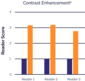
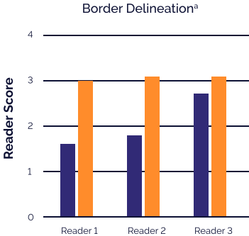
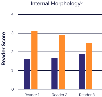
aScoring for degree of contrast enhancement and assessment of border delineation: 1=none, 2=moderate, 3=good, 4=excellent.2
bScoring for internal morphology of lesions: 1=poor, 2=moderate, 3=good, 4=excellent.2
The most common adverse reactions (≥0.65%) reported in clinical trials with Ferabright™ as a contrast agent were nausea, pruritus, constipation, headache, diarrhea, increased blood pressure, bleeding, hyperpigmentation, vein injury, taste alteration, burning/tingling sensation with injection, red sclera, allergic rhinitis, back pain, vomiting, and increased alanine aminotransferase (ALT).1

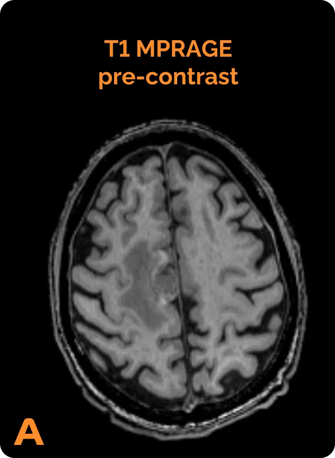
Pre-contrast and Ferabright™- enhanced MRI images of a patient with an intra-axial malignant brain tumor
(A) Pre-contrast MRI. (B) MRI 24 hours after Ferabright™ administration.


Visualization With Ferabright™ Was Comparable to GBCAs1

Lesion visualization scores were similar to those of comparator GBCAs

A similar number of lesions were visualized per patient with each contrast agent

Contrast appearance (lesion size, intensity, and pattern) was sometimes different vs GBCAsc
cIn a side-by-side comparison of post-contrast T1-weighted images, some patients were rated as having larger or smaller size, greater or lesser intensity, and different pattern of lesion contrast between Ferabright™ and gadolinium-based contrast.1